We’re Stronger Together
With your help, we can make ambitious innovations in clinical care and education for our community.
The Interventional Cardiology Program at the International Heart Institute offers comprehensive diagnostic and therapeutic options for non-surgical treatments of cardiac and vascular conditions. Our interventional cardiologists are experts in:
Our cardiovascular specialists work collaboratively with cardiothoracic and vascular surgeons, electro-physiologists, and congenital and heart failure experts. This ensures superior individualized care is provided to every patient to promote health and a better quality of life.
Cardiac catheterization is used to find problems with the heart arteries or measure pres-sures within the heart. It uses x-rays and catheters that are guided via the arteries or veins into the heart to find and treat issues with blood flow. The procedure uses a special contrast dye injected via the catheters to show the anatomy of the heart and vessels. Our specialists access the arteries through the groin or the wrist, which has a lower risk of bleeding and enables patients to walk immediately after catheterization. The procedure typically takes from one to two hours and most patients go home the same day.

Diagnostic Cardiac Catheterization
This procedure is used to measure pressures in the heart chambers and assess the heart valves. It is also used to measure the amount of oxygen in the blood in different areas of the heart. This provides important information about overall heart function, identifies abnormal flows, and measures the pressure and flow in the lungs.
This test is used to clarify if a narrowed section of a coronary artery is likely to cause problems in the future. The catheter is attached to a sensor that measures the pressure in the artery above and below the narrowed area and can determine if a blockage is severe enough to need treatment.

During a catheterization, our specialists can also measure blood pressures and take motion images in the heart chambers to tell if the heart valves are working properly. If you have been diagnosed with heart valve disease, please visit our Structural and Valvular Heart Disease webpage for more details.
This tool uses high-frequency sound waves to create a real-time image of the coronary arteries and identify blockages, as well as allowing our specialists to measure the size of the artery.
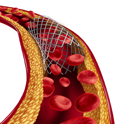
Percutaneous Transcatheter Coronary Angioplasty (PTCA) treats blockages within a vessel and enables a specialist to implant a thin metal mesh, called a stent, to keep the artery open. It involves inserting a thin wire with a deflated balloon on the end into the artery where the blockage is located. The balloon is then inflated to open the vessel. Next, the stent is placed in the area of the blockage to support the artery and keep it open. Most stents contain medication to prevent the artery from closing back up. Blood thinners also help prevent a recurrence of the blockage.

PTCA
Heavily calcified arteries, which prevent a balloon or stent from crossing and being expanded appropriately, can be treated through rotational atherectomy. During the procedure, a high-speed burr is used to grind away calcifications and a stent is inserted into the artery to keep it open.
Chronic total occlusions (CTOs) are complete blockages in the vessels older than 3 months. A specialized skill and set of equipment is needed to open these vessels safely to provide relief from chest pain or other symptoms.
In addition, specialized equipment at the Heart & Vascular Institute can manage more complex or high-risk procedures. These include placing an Impella: a percutaneous Left Ventricular Assist Device (LVAD) or a support pump when a patient’s heart function is very weak or requires support during complex procedures. The pumps can be placed through the leg artery into the heart and usually does not require a surgical procedure.
In this procedure, small samples of heart tissue are obtained to check for signs of rejection in transplanted hearts, to help in diagnosing cardiac disease. The catheter with a small pincer at the tip is introduced through a vein and the samples are obtained. The patient is usually discharged the same day.
During this diagnostic procedure, a catheter, which is guided up the leg vein, carries an ultrasound sensor to provide an image of the heart's internal structure without the need to intubate the patient to place a ultrasound catheter down the throat. ICE is often used for guidance when devices or balloons are placed in the heart. With ICE guidance, some procedures can be performed with the patient awake and discharged the same day.
In a peripheral angiogram, specialists check for problems in the blood vessels in other areas of the body. The device is a high-flow suction catheter which is advanced from the leg or neck veins along the vessel and into the heart to suction out large clots or masses.
Cardiac catheterization is used to find problems with the heart arteries or measure pres-sures within the heart. It uses x-rays and catheters that are guided via the arteries or veins into the heart to find and treat issues with blood flow. The procedure uses a special contrast dye injected via the catheters to show the anatomy of the heart and vessels. Our specialists access the arteries through the groin or the wrist, which has a lower risk of bleeding and enables patients to walk immediately after catheterization. The procedure typically takes from one to two hours and most patients go home the same day.

Diagnostic Cardiac Catheterization
This procedure is used to measure pressures in the heart chambers and assess the heart valves. It is also used to measure the amount of oxygen in the blood in different areas of the heart. This provides important information about overall heart function, identifies abnormal flows, and measures the pressure and flow in the lungs.
This test is used to clarify if a narrowed section of a coronary artery is likely to cause problems in the future. The catheter is attached to a sensor that measures the pressure in the artery above and below the narrowed area and can determine if a blockage is severe enough to need treatment.

During a catheterization, our specialists can also measure blood pressures and take motion images in the heart chambers to tell if the heart valves are working properly. If you have been diagnosed with heart valve disease, please visit our Structural and Valvular Heart Disease webpage for more details.
This tool uses high-frequency sound waves to create a real-time image of the coronary arteries and identify blockages, as well as allowing our specialists to measure the size of the artery.
Percutaneous Transcatheter Coronary Angioplasty (PTCA) treats blockages within a vessel and enables a specialist to implant a thin metal mesh, called a stent, to keep the artery open. It involves inserting a thin wire with a deflated balloon on the end into the artery where the blockage is located. The balloon is then inflated to open the vessel. Next, the stent is placed in the area of the blockage to support the artery and keep it open. Most stents contain medication to prevent the artery from closing back up. Blood thinners also help prevent a recurrence of the blockage.

PTCA
Heavily calcified arteries, which prevent a balloon or stent from crossing and being expanded appropriately, can be treated through rotational atherectomy. During the procedure, a high-speed burr is used to grind away calcifications and a stent is inserted into the artery to keep it open.
Chronic total occlusions (CTOs) are complete blockages in the vessels older than 3 months. A specialized skill and set of equipment is needed to open these vessels safely to provide relief from chest pain or other symptoms.
In addition, specialized equipment at the Heart & Vascular Institute can manage more complex or high-risk procedures. These include placing an Impella: a percutaneous Left Ventricular Assist Device (LVAD) or a support pump when a patient’s heart function is very weak or requires support during complex procedures. The pumps can be placed through the leg artery into the heart and usually does not require a surgical procedure.
In this procedure, small samples of heart tissue are obtained to check for signs of rejection in transplanted hearts, to help in diagnosing cardiac disease. The catheter with a small pincer at the tip is introduced through a vein and the samples are obtained. The patient is usually discharged the same day.
During this diagnostic procedure, a catheter, which is guided up the leg vein, carries an ultrasound sensor to provide an image of the heart's internal structure without the need to intubate the patient to place a ultrasound catheter down the throat. ICE is often used for guidance when devices or balloons are placed in the heart. With ICE guidance, some procedures can be performed with the patient awake and discharged the same day.
In a peripheral angiogram, specialists check for problems in the blood vessels in other areas of the body. The device is a high-flow suction catheter which is advanced from the leg or neck veins along the vessel and into the heart to suction out large clots or masses.
With your help, we can make ambitious innovations in clinical care and education for our community.